
Mackay School of Earth Science and Engineering
Microbeam Laboratory
University of Nevada, Reno
Examples of Applications
Ore Deposits
BSE and EDS Elemental Maps of Silver-bearing Pyrite from a Carlin-style Gold-Silver Deposit
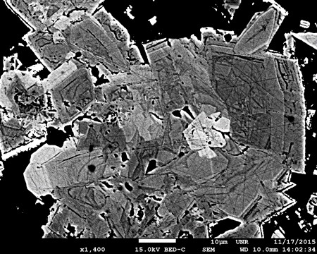
Backscatter electron image (BSE) of pyrite from the Cove Carlin-style gold-silver deposit in Nevada. The variation of brightness is a function of arsenic substituting for sulfur in the pyrite crystal structure. Note the complex growth history of the pyrite. Image collected from a polished thin section using the BSE detector on the JEOL 7100FT FESEM.
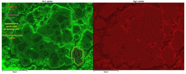
Energy dispersive spectroscopy (EDS) elemental maps showing distribution of As (left) and Ag (right) from the same pyrite from Cove deposit. Brighter zones represent areas with higher concentrations of As (left) and Ag (right). Highest As and Ag occur in late outermost rims on the grains. Brighter zones (right) represent area with higher concentrations of Ag. Cove is one of the few Carlin-style deposits with documented Ag in ore-stage pyrite. Prior to this image, Ag was documented post analyses of pyrites from Cove using SIMS, an expensive method available in only a few labs. The image demonstrates Ag is detectable with the Oxford EDS system on the JEOL 7100FT FESEM.
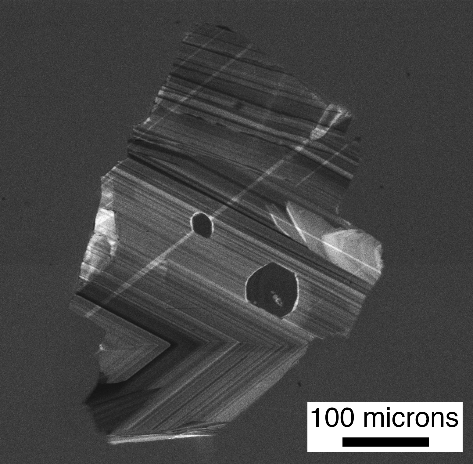
CL image of epithermal gold-bearing quartz vein
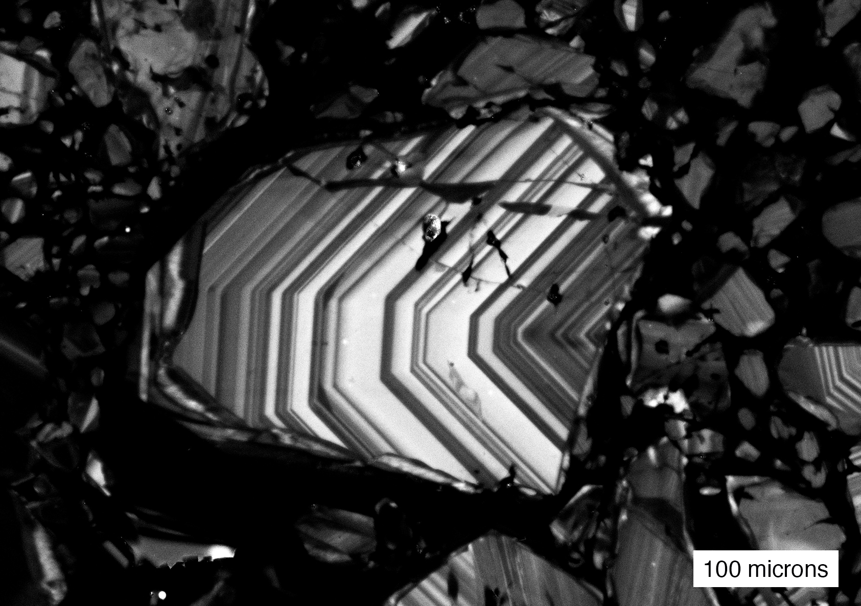
Cathodoluminescence (CL) image of a laminated syntaxial quartz vein containing free gold, Bi minerals, fluorite, and tourmaline from a gold prospect in the Union District, NV. Sub-micron spatial resolution achieved through CL is now considered standard in understanding ore and fluid inclusions and paragenesis in vein deposits. For example, in fluid inclusion studies aimed at understanding the temperature, pressure, and salinities of ore fluids, one needs to focus on fluid inclusions in the generation of quartz that hosts the ore minerals. Image acquired from a polished thin section using the Deben panchromatic CL detector on the JEOL 7100FT FESEM.
Industrial Minerals
Secondary Electron images of Lithium-bearing Clays
 |
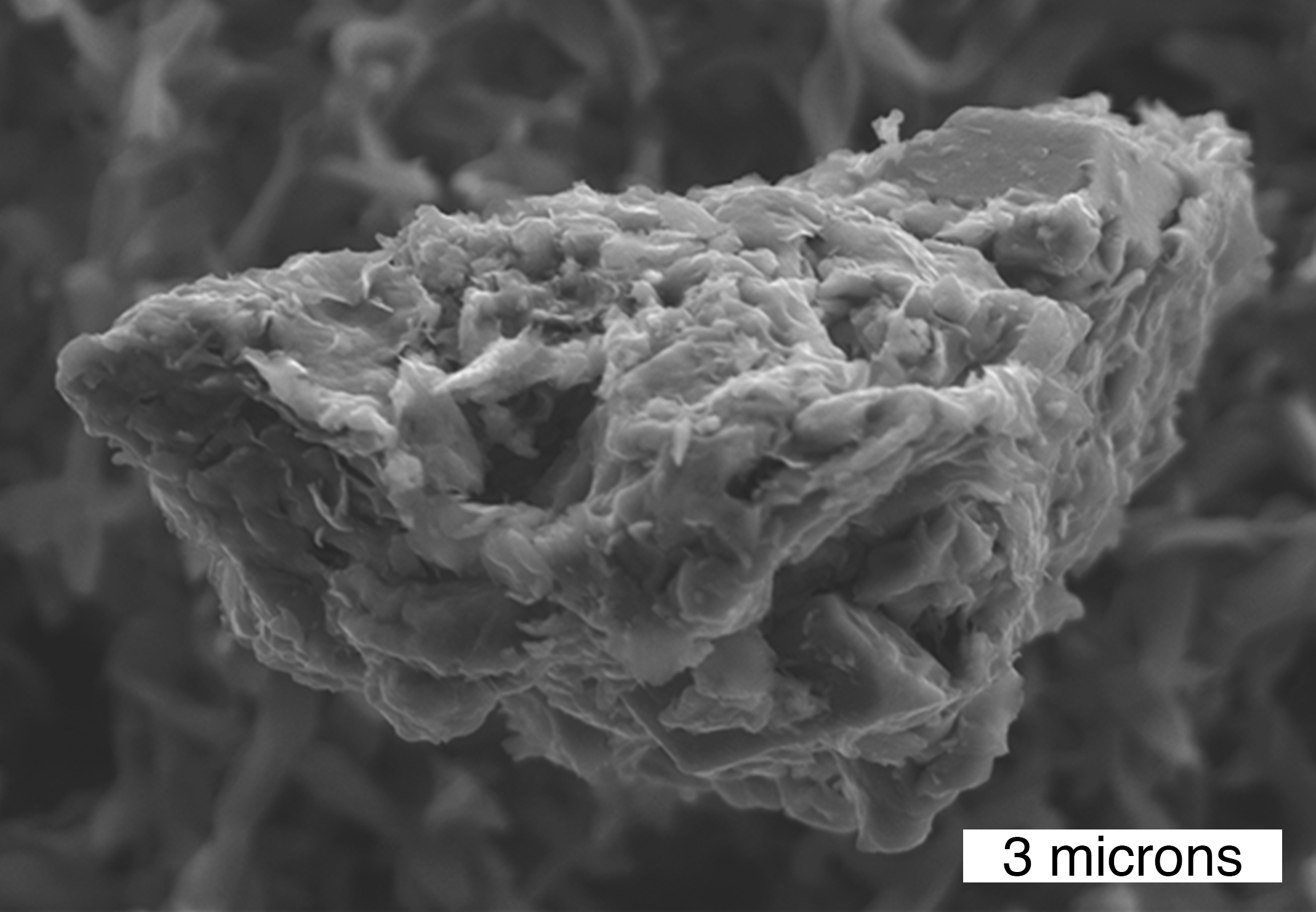 |
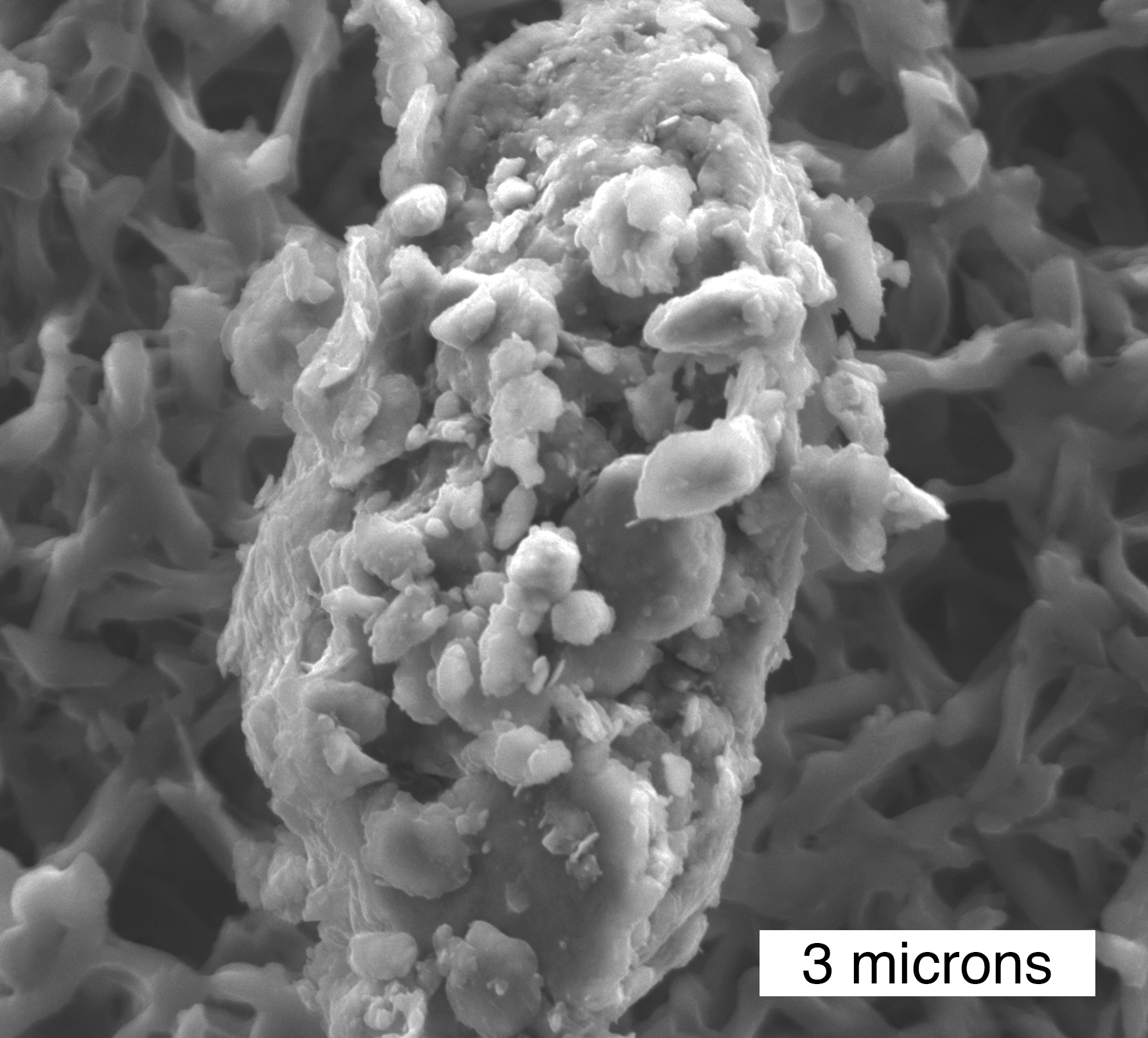
Secondary electron (SE) image of lithium bearing clay from a lithium deposit in Nevada. The lithium clay is not only a potential source of lithium, but the clay is used as an additive to drilling mud. The potential industrial applications of clays are dependent upon the physical properties of the clay (e.g., size, morphology, chemistry). In this case, a private firm utilized the JEOL 7100FT FESEM to document the morphology of the clay particles.
Petrology and Tectonics
Phases in Ultrahigh-pressure Eclogites
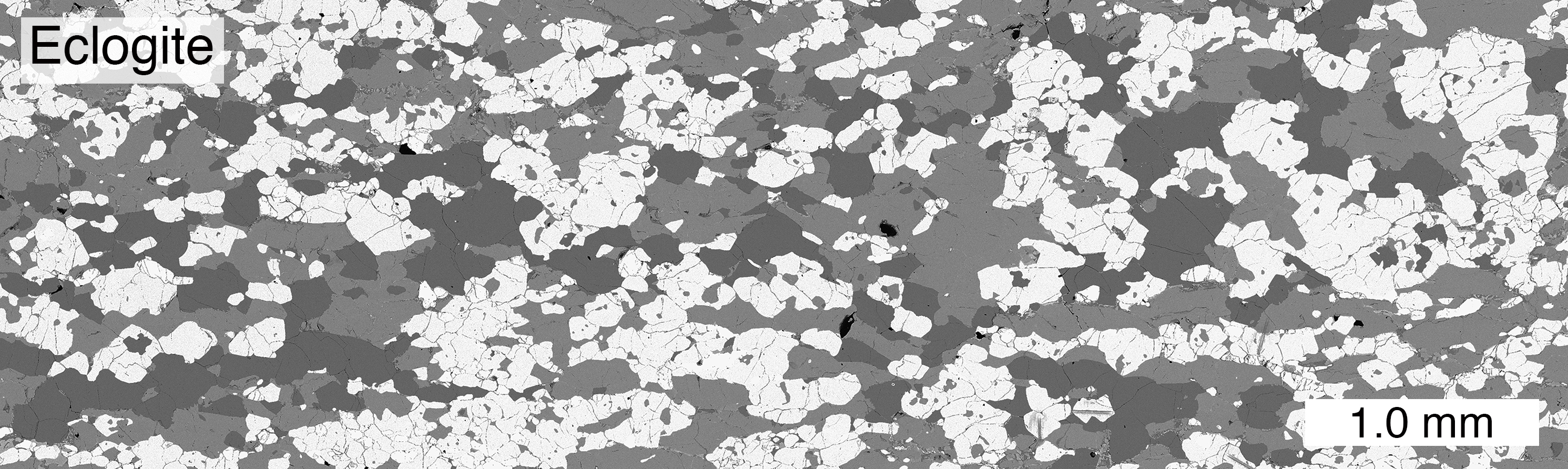
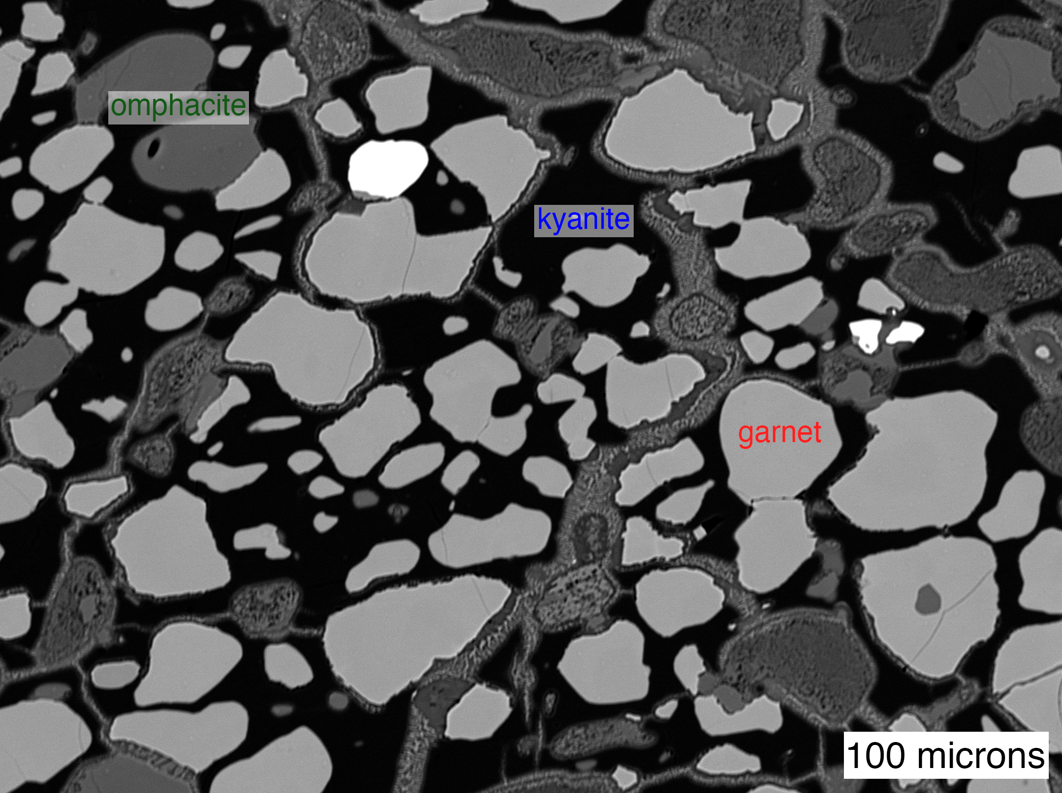
Top: Large scale backscatter electron (BSE) scan from an ultrahigh-pressure eclogite showing distribution and proportions of major phases throughout the thin section: quartz-dark grey, omphacite-grey, garnet-light grey. Bottom: BSE image of a kyanite eclogite showing a kyanite poikoloblast that contains garnet and omphacite inclusions. Note the formation of symplectite at the expense of the peak phases during retrogression in the lower–middle crust. Images were generated with the BSE system on the JEOL 7100FT FESEM (courtesy Stacia Gordon and Joel DesOrmeau).
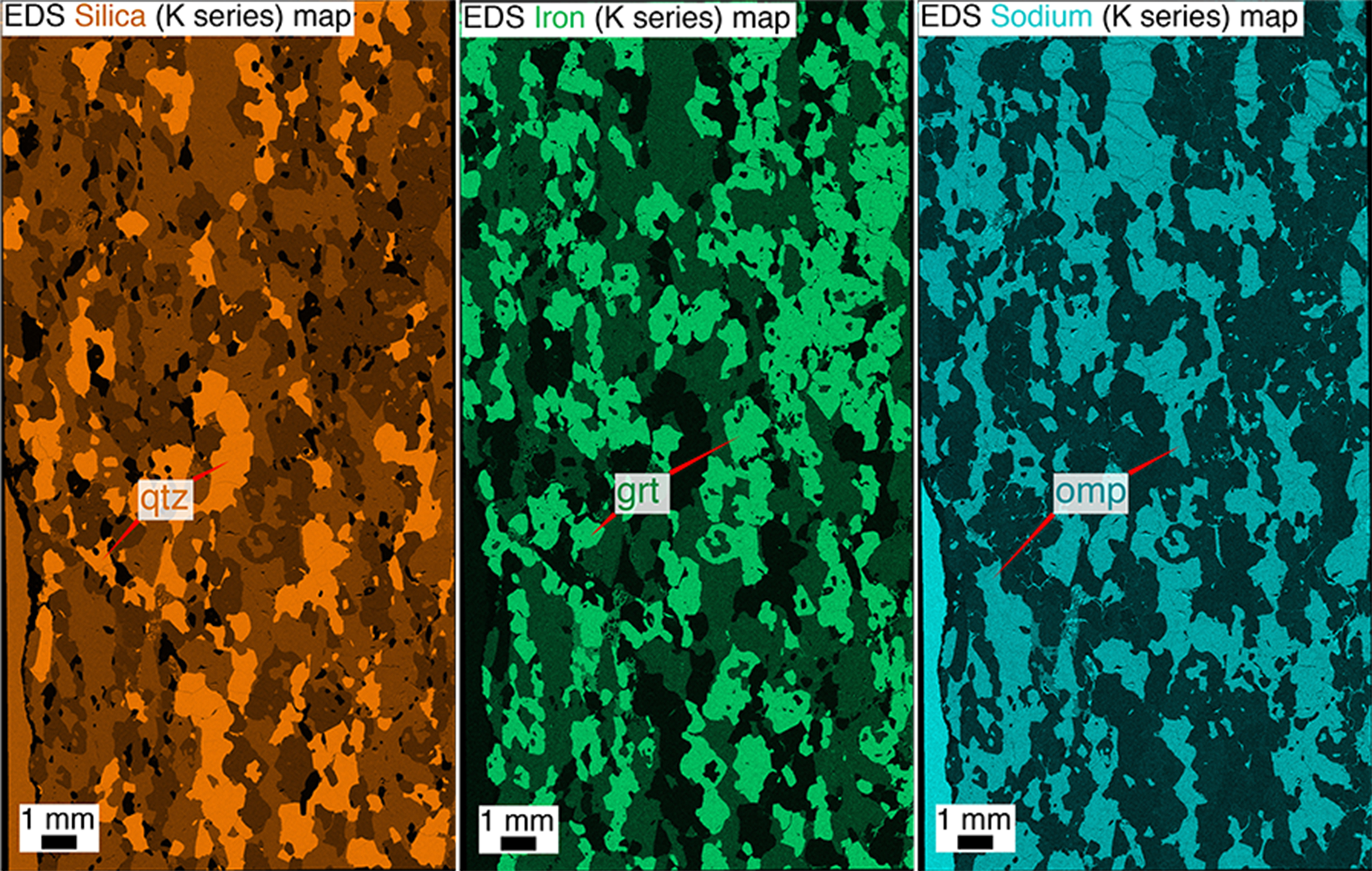
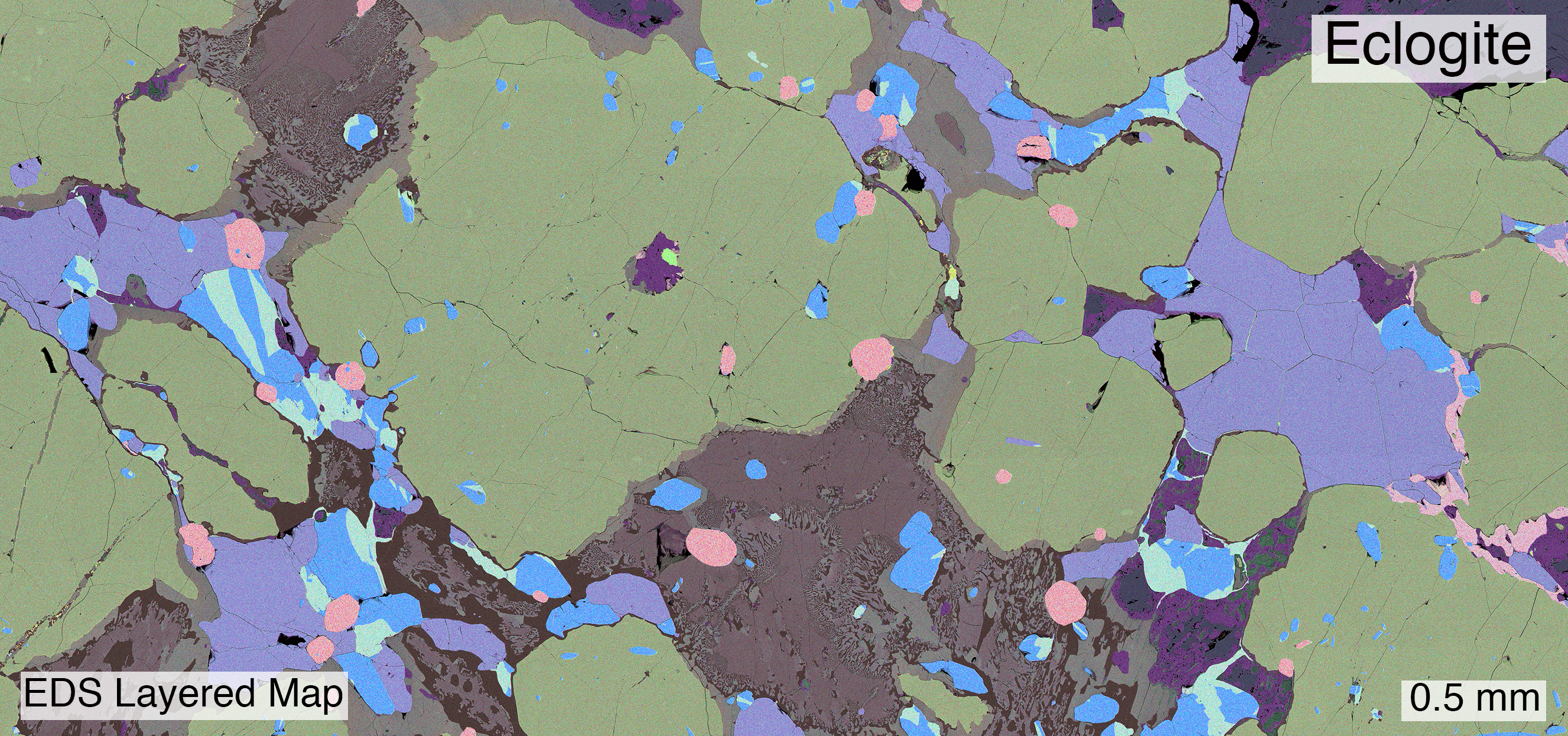
Top: Large scale EDS maps of silica, iron, and sodium K series from an ultrahigh-pressure eclogite showing phase distribution and proportions throughout the thin section: (left) quartz-orange, (middle) garnet-green, and (right) omphacite-blue. Bottom: Layered EDS maps of high-pressure eclogite showing textural relationships of accessory mineral (zircon-pink and rutile-blue) growth and breakdown of peak phases. Individual element maps generated with the Oxford EDS detector on the JEOL 7100FT FESEM and layered map compiled with Oxford Aztec software (courtesy Stacia Gordon and Joel DesOrmeau).
Orthoamphibole Gneiss
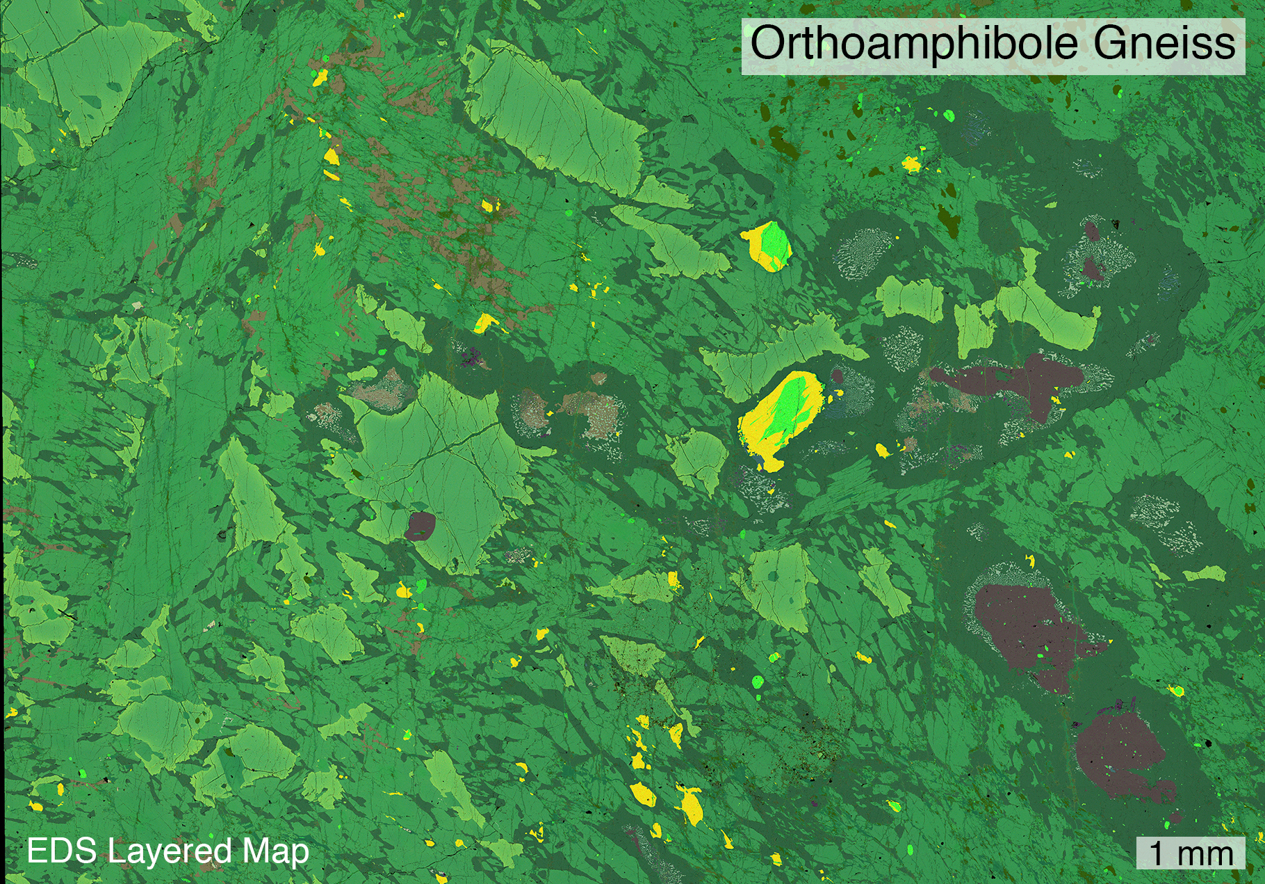
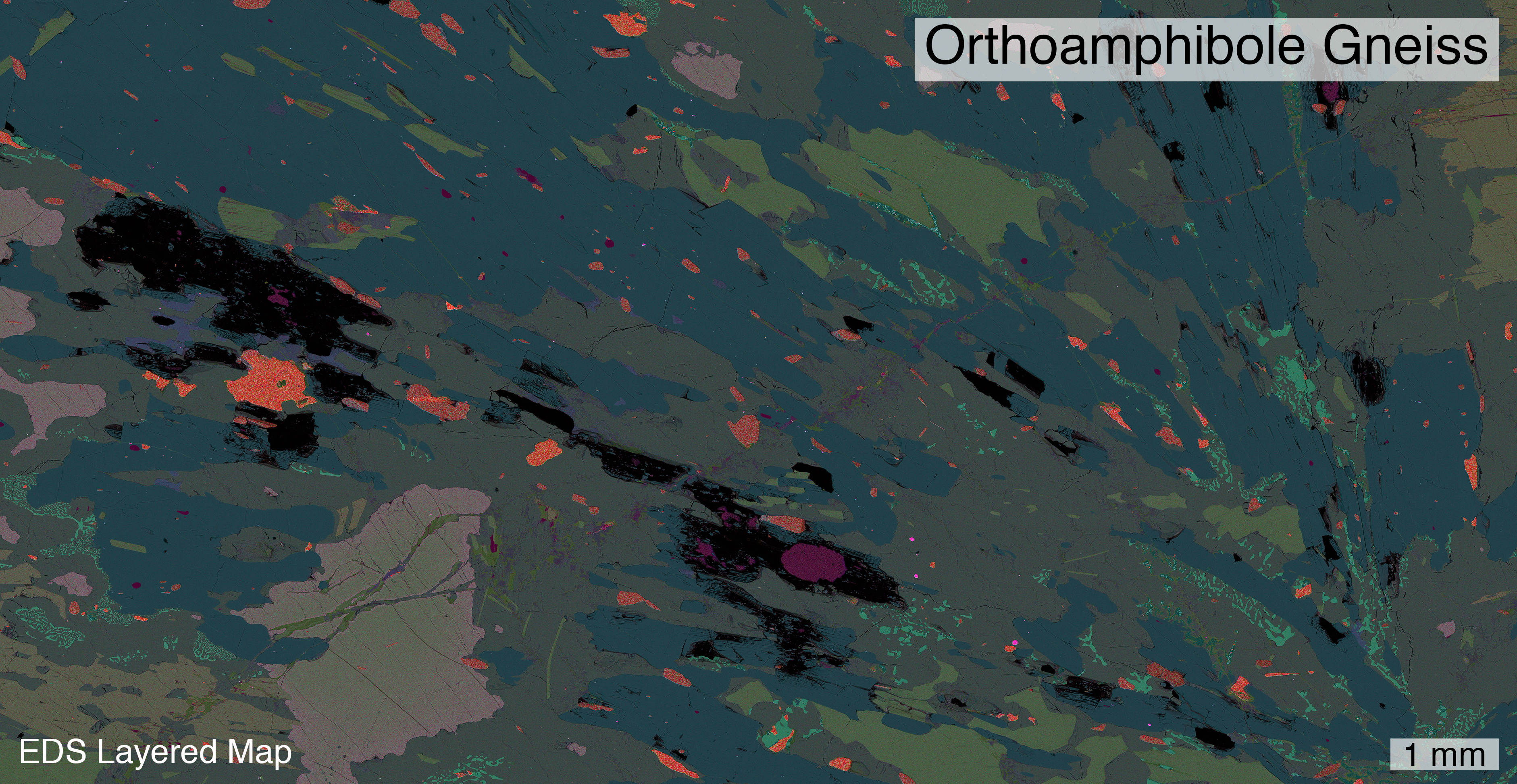
Layered EDS maps of orthoamphibole gneisses showing complex textural relationships between the main matrix phases of gedrite, garnet, kyanite, and symplecite of spinel, sapphirine, plagioclase, and cordierite. Individual element maps generated with the Oxford EDS detector on the JEOL 7100FT FESEM and compiled with Oxford Aztec software (courtesy Stacia Gordon).
CL Images of Accessory Minerals
 |
 |
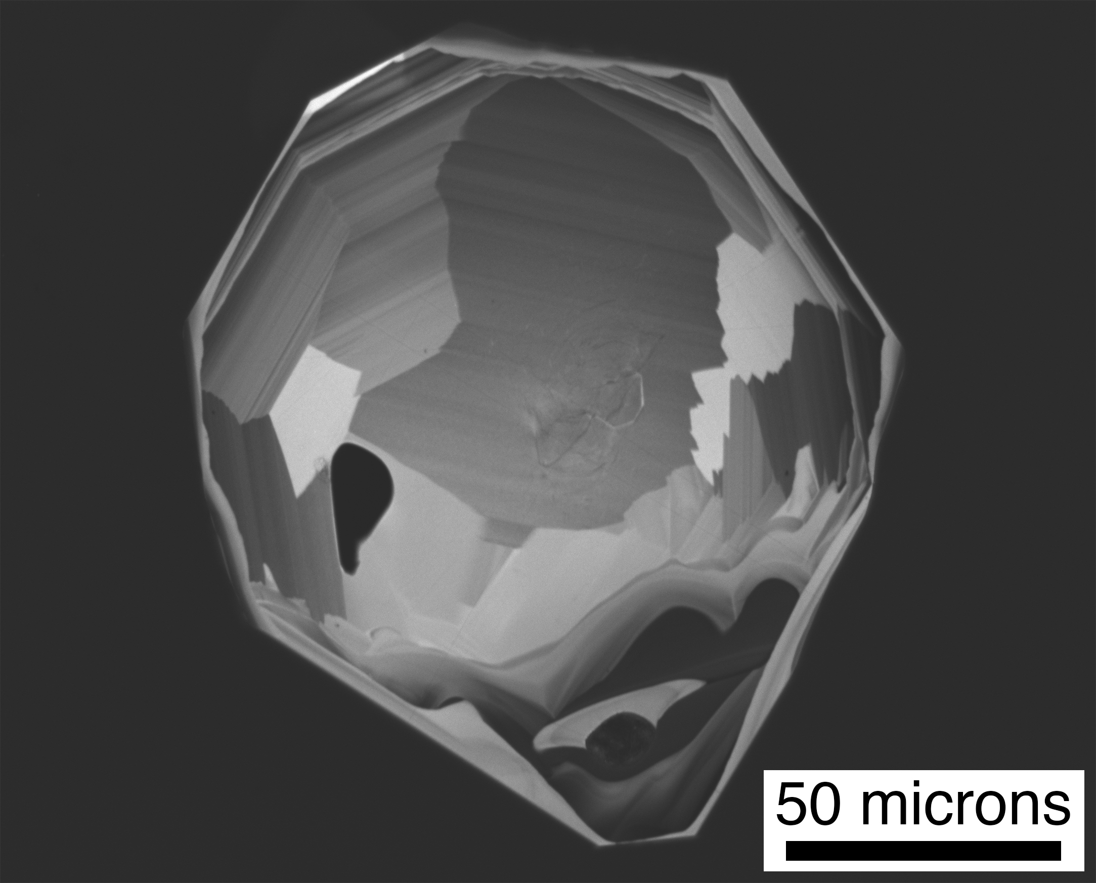
Cathodoluminescence images showing complex accessory mineral growth history: (left) sector and oscillatory zoning preserved within zircon (ZrSiO4) and (right) oscillatory zoning showing alternating thickness of zones preserved within Baddeleyite (ZrO2). Images taken with the Deben panchromatic CL detector on the JEOL 7100FT FESEM.
Paleontology
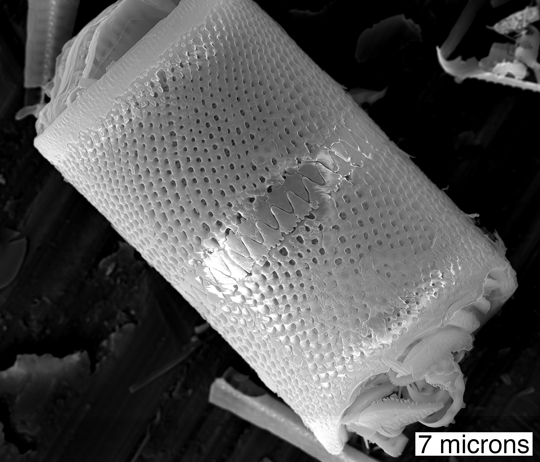
Identification of Diatoms
 |
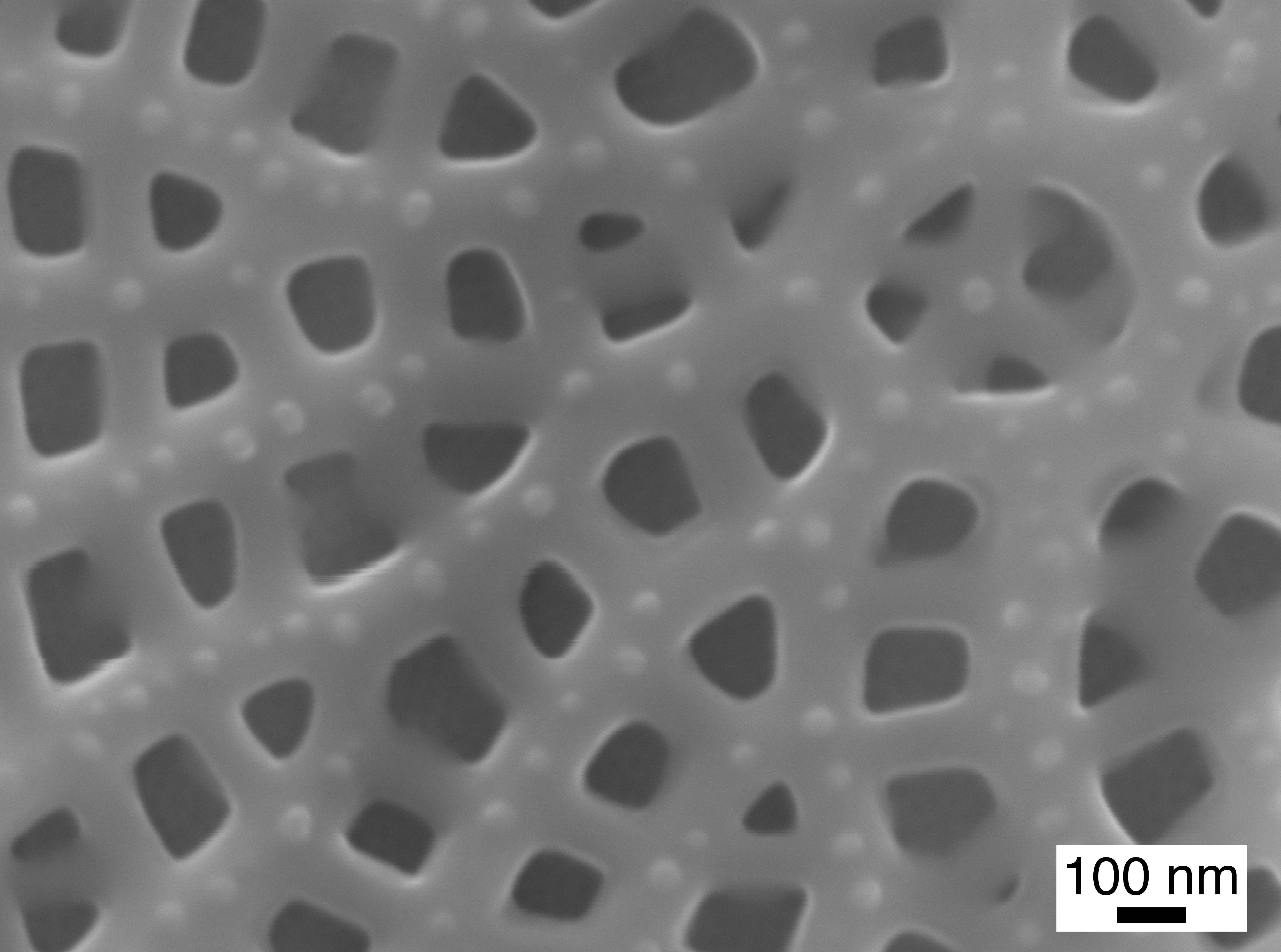 |
Secondary electron (SE) images of diatom specimens showing the high-resolution (ten’s of nanometers; right) capabilities of the JEOL 7100FT FESEM (courtesy Kerry Howard and Dr. Paula Noble).
Engineering and Material Sciences
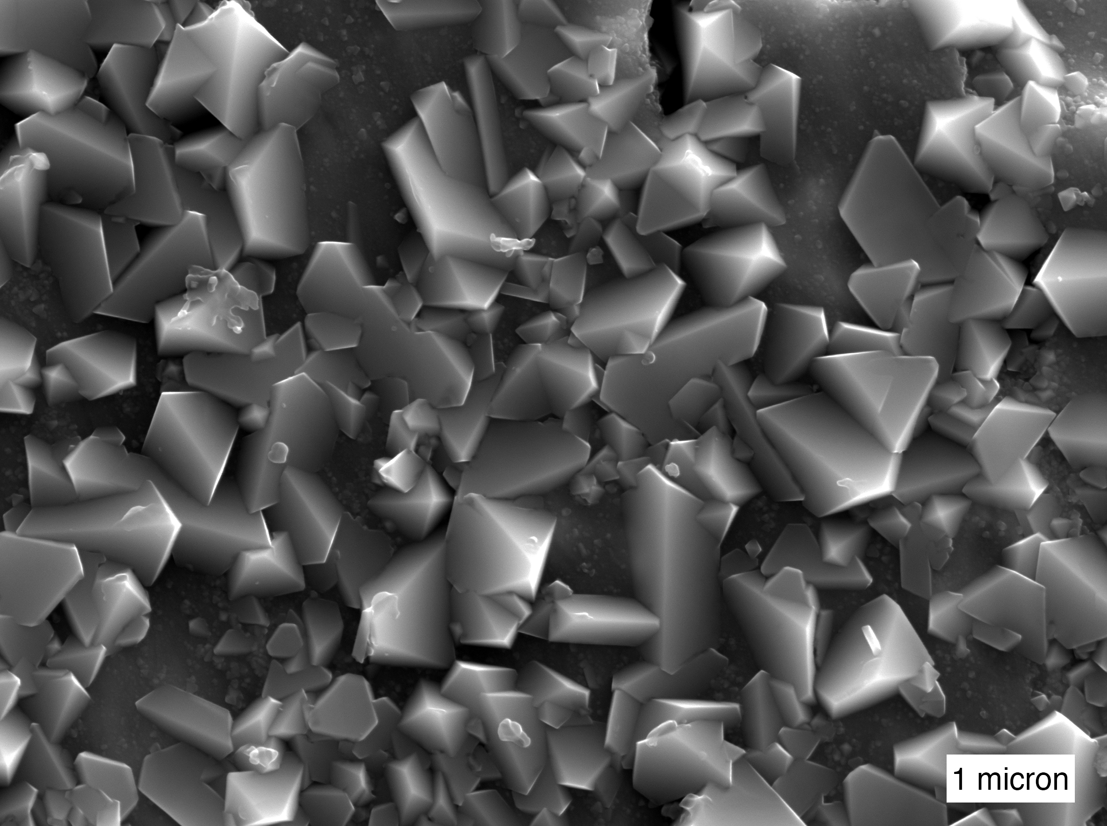
Secondary electron (SE) image of oxide formation on stainless steel. Image taken with the JEOL 7100FT FESEM (courtesy Zachary Karmiol and Dr. Dev Chidambaram).
Microstructural Studies of Magnesium Alloys
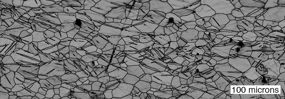
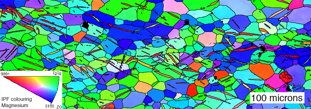
Electron backscatter diffraction (EBSD) band contrast (top) and inverse z-pole (bottom) figures showing crystallographic information of tested samples of rolled magnesium alloy (courtesy Dr. Yanyao Jiang & Duke Culbertson). EBSD allows for characterization of microstructural and textural information (orientation mapping and texture determination, deformation and twinning, grain size measurements and grain boundary analysis, phase identification, strain analysis, and elasticity).
Other applications
Biology (ex., cell studies)
Chemistry (ex., bond microstructure)
Physics
Metallurgy (ex., textural analysis, dynamic recrystallization)
Civil Engineering (ex., texture and failure analysis, dynamic recrystallization)
Forensic science (ex., coupled high-resolution imaging and EDS chemical analysis)
Archeology(ex., high-resolution imaging)
Environmental Science (ex., coupled high-resolution imaging and EDS chemical analysis)
Pharmaceuticals (ex., coupled high-resolution imaging and EDS chemical analysis)
Publications and Theses affiliated with the Microbeam Lab
Culbertson D., and Jiang Y, (submitted manuscript), An experimental study of the orientation effect on fatigue crack propagation in rolled AZ31B magnesium alloy, Materials Science and Engineering (2016).
Sauer, K.B., Gordon, S.M., Miller, R.B., Vervoort, J.D., and Fisher, C.M., (submitted manuscript), Detrital zircon U-Pb and Hf provenance of the Late Mesozoic sedimentary units of the North Cascades continental arc, Washington, Geosphere (2016).
Time-space relationships between sediment-hosted gold mineralization and intrusion-related polymetallic mineralization at Kinsley Mountain, NV, Tyler Hill M.S. Thesis (2016).