Departments, Labs, and Facilities
Departments
Great Basin Science Sample and Records Library (GBSSRL)
Ralph J. Roberts Center for Research in Economic Geology (CREG)
Great Basin Center for Geothermal Energy (GBCGE)
Cartography, GIS, and Publications Support
Administration & Director's Office
Labs and Facilities
Raman spectrometry lab
The Raman spectroscopy laboratory is managed by NBMG. Raman spectroscopy allows for rapid, non-destructive characterization of minerals, fluids, dissolved gasses, and more. Measuring the "Raman shift" of a sample can provide information about the chemical composition, structure, and properties of a sample.
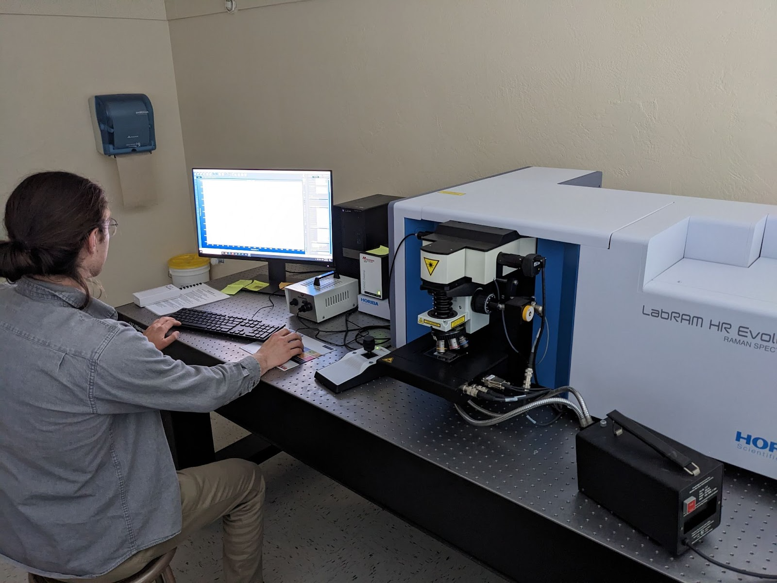
PhD student Dominik Vlaha working in the Raman spectroscopy laboratory.
The lab consists of a Horiba LabRAM HR Evolution spectrometer optimized for 200-2200 nm equipped with an open space confocal microscope with 5 objectives (5x, 10x, 50x, 50x LWD, and 100x), a Marzhauser XY motorized stage, two diffraction gratings (600 and 1800 gr/mm) and a multichannel CCD detector for a wide spectral resolution range. The excitation beam is provided by a frequency doubled Nd:YAG laser (Oxxius, France) at 532 nm with a maximum power of 100 mW.
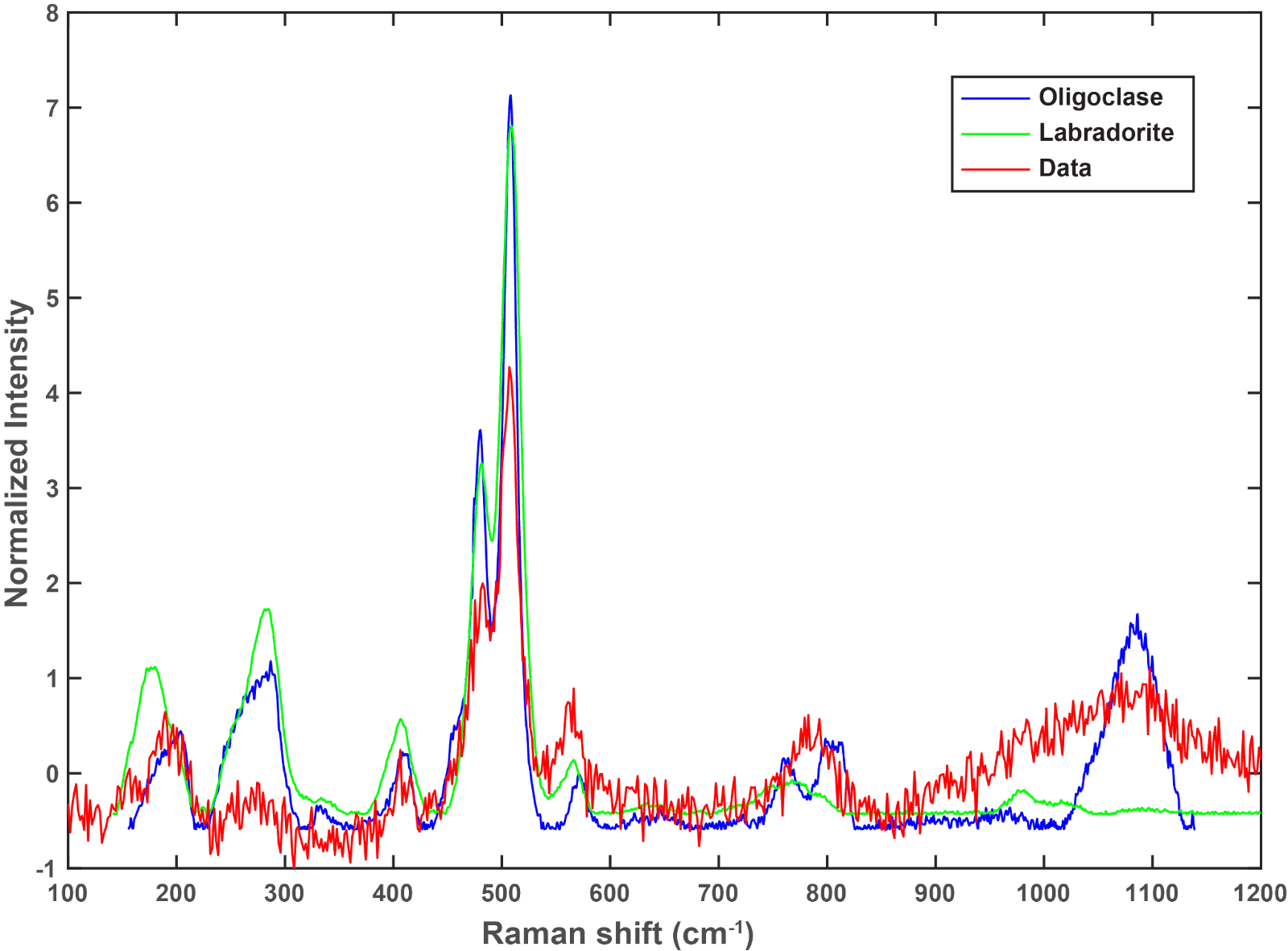
Raman spectroscopy is a non-destructive technique that allows us to recognize mineral structures. In this case, the mineral spectra (red line) can be identified as Labradorite or Oligoclase (data base from RRUFF project https://rruff.info/about/about_general.php).
The lab is primarily used for structural and thermobarometric studies such as Raman Spectroscopy of Carbonaceous Material (RSCM), quartz-inclusion barometry, and mineral identification. For RSCM work, we have a lab-specific calibration curve from the reference standards and data reduction protocols of Lunsdorf et al. (2017).
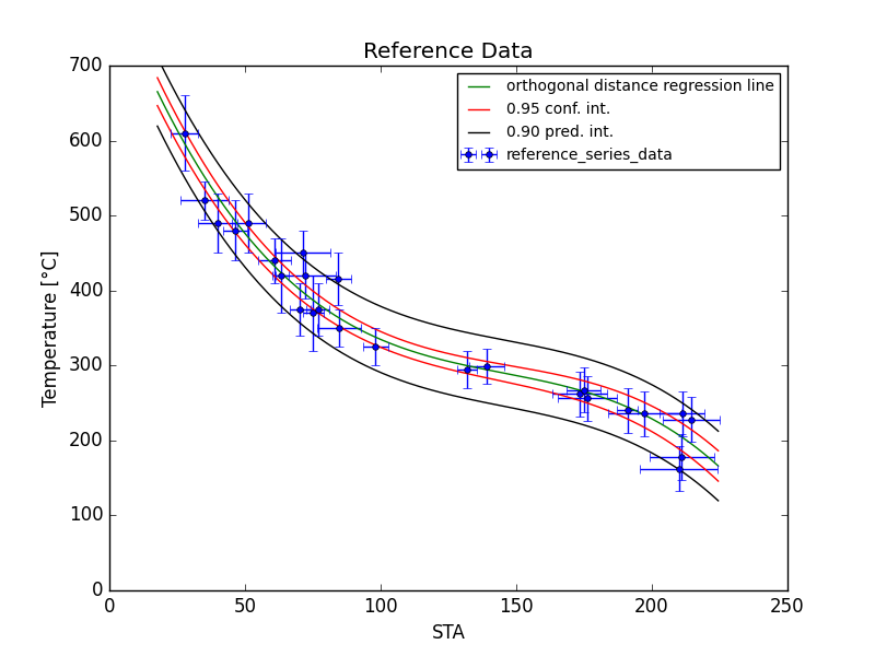
Lab-specific calibration curve for RSCM analyses showing the relationship between scaled total area (STA) and peak temperature using the reference standards and data reduction protocols of Lunsdorf et al. (2017).
Fluid inclusion lab
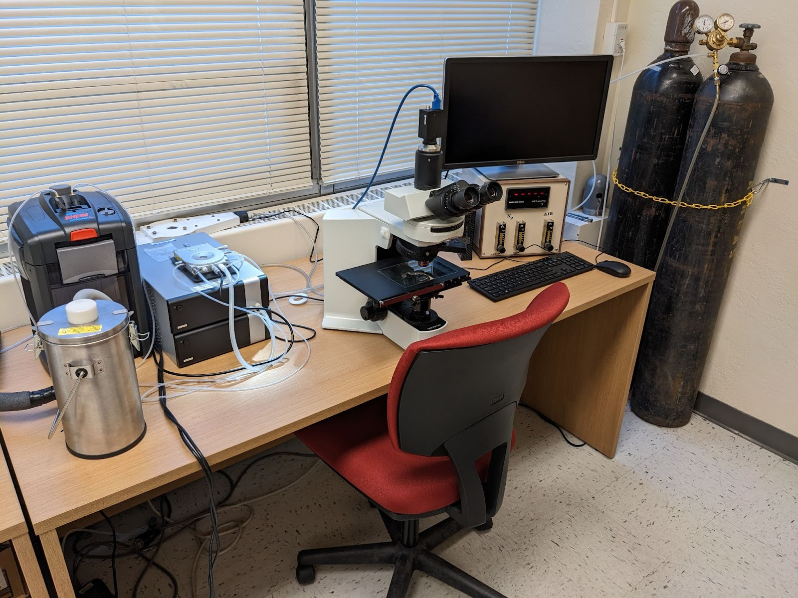
Fluid inclusion freezing-heating system and microscope.
Mineral separation facilities:
There are two rock-sample-preparation laboratories that have equipment for thin-section making, crushing, mineral separation, and powdering. This includes a trim and slab rock saw, water shaking table, sieves, magnetic separator, various heavy liquids, and fume hoods. These labs are primarily used for cutting and trimming thin-section billets, powdering samples for geochemical analysis, and separating minerals for geo-/thermochronology applications including zircon, apatite, sanidine, muscovite, biotite, and hornblende.
Optical microscopes with digital cameras:There are numerous transmitting and reflecting light optical microscopes with camera systems. There are also binocular stereo picking microscopes. The newest microscope camera connects to a powerful computer with Leica LAS X software installed for image analysis, including powerful measuring features.
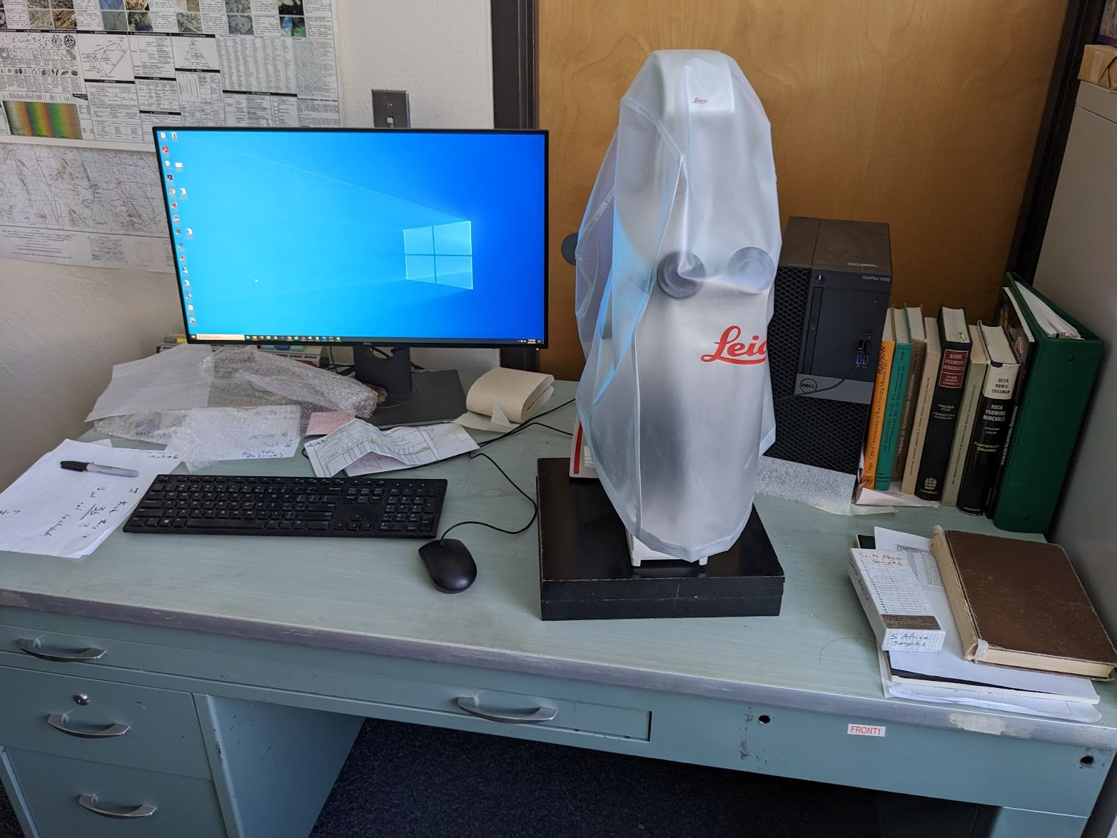
Leica petrographic microscope and computer with imaging software.
Mackay Microbeam Laboratory
Established in 2015, the Mackay School of Earth Science and Engineering Microbeam Laboratory houses two state of the art JEOL scanning electron microscopes (SEM), a field emission SEM and an easy to use portable tungsten-filament SEM. The field emission SEM has an Oxford energy dispersive spectroscopy (EDS) and electron backscatter diffraction (EBSD) systems and a Deben cathodoluminescence (CL) detector. The tungsten-filament SEM has low-vacuum capabilities, which allows for examination of biologic and other moist materials. The lab is transforming research in the Earth Sciences at Mackay and within the College of Science and is being used increasingly by other departments and colleges at UNR. The machine is also utilized by entities outside UNR, fostering cooperation and collaboration among UNR, the city of Reno, greater Nevada, and researchers throughout the world. Our mission is to make the Mackay School of Earth Science and Engineering Microbeam Laboratory the microscopy lab of choice for the western U.S.
Former Labs
Nevada Analytical Laboratory

 Home
Home